

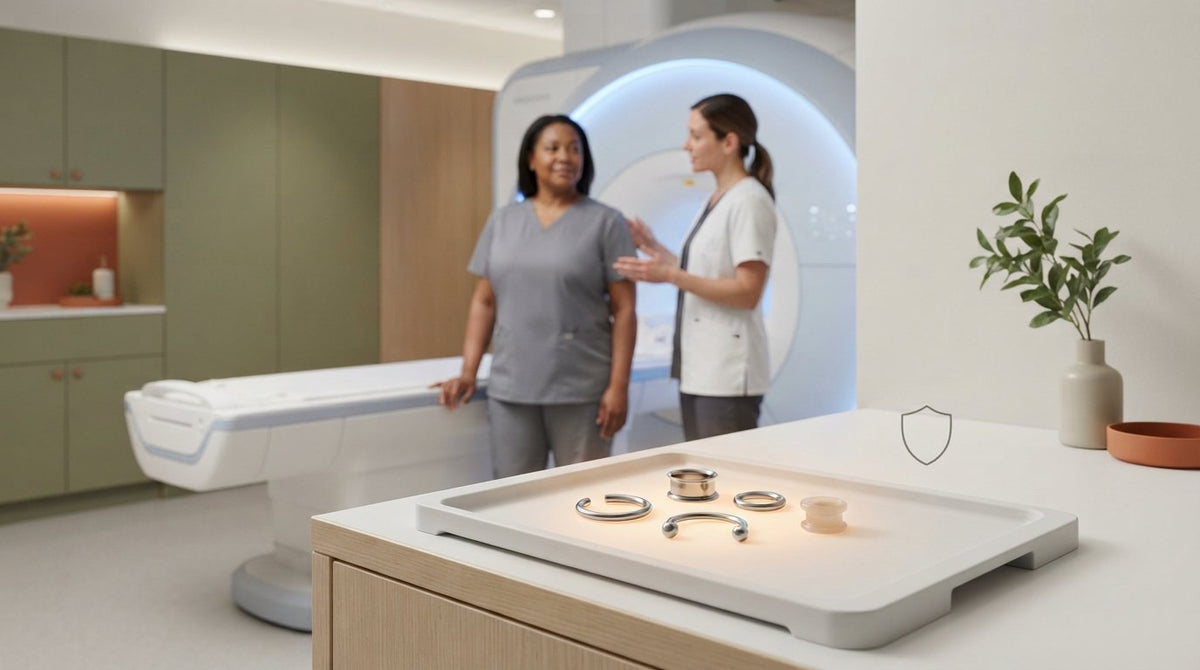
A surgical-steel piercing isn’t automatically safe just because it’s labelled “surgical.” Whether you can get an MRI with surgical steel piercings depends on the jewelry’s material, location, and the area being scanned. Metallic body jewelry should be removed before entering the MRI environment, and surgical steel is often required to come out for safety and image clarity.
If you have stretched lobes, you may worry that removing plugs or tunnels could cause the piercing to shrink or close. That’s a real consideration, but leaving metal in place can also pose risks: some jewelry may move, heat up, or affect the images. Tell your healthcare team about every piercing and what you know about its material before the scan. If a piece can’t be removed, the MRI team will assess its location and material and explain relevant precautions.
This guide explains why surgical steel can be a concern, how titanium differs, and what to consider if a piercing needs to stay open. You’ll also learn how stretched-lobe jewelry fits into MRI preparation and when a temporary nonmetallic retainer may be an option.
Key Takeaways
- Surgical steel isn’t automatically MRI-safe; material, piercing location, and the area being scanned all matter.
- Metal jewelry may move, heat up, or affect image quality, so tell the imaging team about every piercing before the scan.
- Nonmetallic retainers can help keep a piercing open, but the MRI team needs to know what material is in place.
- For stretched lobes, plan ahead: removing plugs or tunnels may affect the piercing, so discuss options before your appointment.
- Ask a healthcare professional about MRI precautions and a piercer about jewelry material or removal when you need support preparing.
Heating, magnetic pull, and image distortion risks
Surgical steel isn’t automatically nonmagnetic or safe for an MRI. Some surgical stainless steel, including grade 316LVM, may show magnetic pull in an MRI despite being considered low-magnetic or nonmagnetic for everyday purposes. The alloy and the scan both matter.
Ferromagnetic metals can respond to the scanner’s strong magnetic field. Iron, cobalt, and nickel are examples of ferromagnetic materials that can cause problems. Jewelry containing ferrous metal may move, become displaced, or act as a projectile in the MRI environment. Movement can cause discomfort, and severe cases can result in injury.
Conductive metal presents another concern: radiofrequency energy during an MRI can heat jewelry and raise its temperature. A hot ring, barbell, or plug may burn the skin it touches. The risk depends partly on the jewelry’s material, shape, and position, so its appearance alone cannot establish whether it’s safe for an MRI.
Image interference matters, too. Metal can distort the magnetic field near the jewelry, creating artifacts that obscure details in the scan. A piercing near the area being imaged may interfere more with interpretation than one farther away. For example, facial jewelry can affect head or face images, even when image quality, rather than movement, is the main concern.
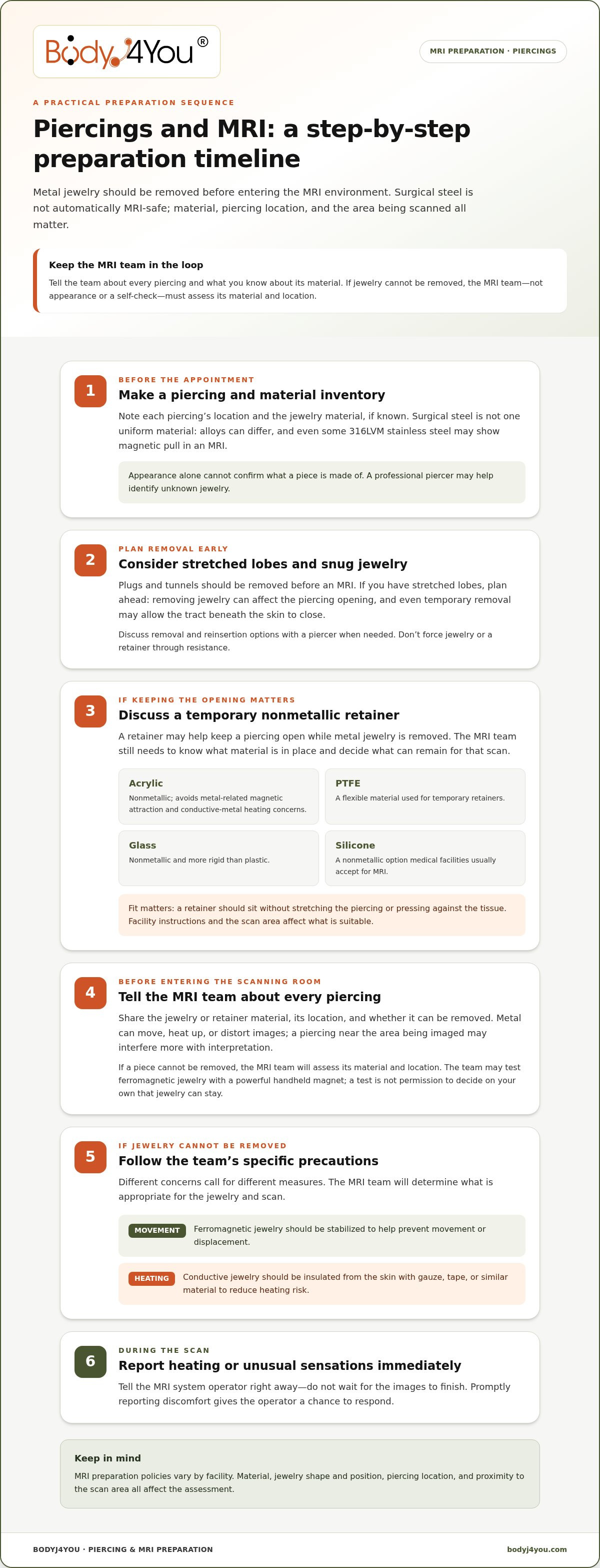
If jewelry can’t be removed, tell the MRI technologist before entering the scanning room. The team may assess its material and location, and ferromagnetic jewelry may be tested with a powerful handheld magnet. A magnet test is not a reason to conceal a piercing or decide on your own that it can stay in place. The imaging team needs complete details to assess the situation.
When removal isn’t possible, stabilization and insulation address different concerns. Ferromagnetic jewelry should be stabilized to help prevent movement or displacement. Conductive jewelry should be wrapped with gauze, tape, or similar material to insulate it from the skin and reduce the risk of heating. Yale Medicine may ask patients to place an ice pack over a piercing during an MRI to keep metal cool.
Tell the MRI system operator immediately if you feel heating or any unusual sensation during the scan. Don’t wait for the images to finish. Reporting discomfort promptly gives the operator a chance to respond. Surgical steel isn’t one uniform material: its alloy, magnetic response, conductivity, location, and proximity to the scan area all affect safety and image quality.
Non-metallic retainers and material choices
A temporary retainer can help keep a piercing open while metal jewelry is removed for an MRI. The key distinction is material: acrylic, PTFE, glass, and silicone are nonmetallic options. They don’t create the same metal-related concerns, but the imaging team still needs to know what’s in place before the scan.
Acrylic is nonmetallic and doesn’t pose risks from magnetic attraction or conductive-metal heating. PTFE is a flexible material used for temporary retainers. Glass is also nonmetallic, but feels more rigid than plastic. Glass and silicone retainers are options medical facilities usually accept for MRI, though the imaging team determines what can remain during a particular scan.
- Acrylic: A nonmetallic option without the MRI risks associated with metal jewelry.
- PTFE: A flexible material used for temporary retainers.
- Glass: A nonmetallic retainer that is more rigid than plastic.
- Silicone: A nonmetallic option that medical facilities usually accept for MRI.
These materials aren’t interchangeable for every piercing. For example, a rigid glass retainer may feel different from a flexible PTFE retainer, while the piercing’s location affects which shape can sit comfortably. The opening’s size and the jewelry’s fit matter, too. A retainer should fit the piercing without stretching or pressing against the tissue.
Identifying the material is a practical first step. If you don’t know what a piece is made from, a professional piercer may help identify it and discuss a temporary nonmetallic retainer. That distinction matters because a metal-coloured finish doesn’t establish the underlying material. Tell the MRI team the retainer’s material, location, and whether it can be removed.
Plan for removal and reinsertion, too. Even temporary removal can allow the piercing tract beneath the skin to close, so a spacer may be useful. A professional piercer may assist with removal and reinsertion, especially if the piercing is difficult to reach or the jewelry is snug. Don’t force a retainer through resistance or use a size that changes the fit.
Think of the retainer as a temporary bridge, not permission to keep jewelry in automatically. MRI preparation policies vary between facilities, and the scan area can affect image quality. For example, a retainer near the body part being imaged may require a different decision from one farther away. Share the material and placement details before the appointment so the imaging team can account for them.
For stretched lobes, discuss plugs or tunnels separately from smaller piercing jewelry. Heavy plugs and tunnels should be removed before an MRI, and a nonmetallic retainer may help preserve the piercing opening. The right option depends on fit, material, and the facility’s MRI instructions. Planning ahead can reduce last-minute changes while keeping safety at the centre.
Preparing for an MRI with piercings
Yes, instructions can differ depending on the piercing’s location and the scan area. A facial piercing may affect head or face images, while the same jewelry elsewhere may pose a different concern. Tell the imaging team where each piercing is and what it’s made from.
Piercing Preparation Before Scanning
MRI preparation means identifying every piercing and following the imaging team’s instructions before entering the scanner room. Tell your physician, imaging scheduler, and technologist about jewelry and other items on or in your body that weren’t present at birth. Metallic body jewelry should be removed before entering the MRI environment. Planning early gives the team time to address piercings that may be difficult to remove.
Before your appointment, gather details that help identify the jewelry. If you have a mill certificate documenting its alloy, bring it and show it to the imaging team. Mention recent piercings, too. If a piercing is less than 6 to 8 weeks old, contact radiology ahead of the MRI. A newer piercing may need special planning if jewelry removal risks the opening closing.
Body Area and Scan Location
The scan location matters because metal can interfere with images near the jewelry. For example, facial piercings may distort head or face images, while jewelry farther from the scanned area may affect image quality differently. Location also helps the MRI team assess how close the metal is to the imaging area. Share the exact piercing site, not just the number of piercings.
Use a clear inventory to avoid overlooking less visible jewelry:
- List each piercing’s body location and jewelry material.
- Include stretched lobes, plugs, and tunnels.
- Remove heavy plugs and tunnels before the MRI.
- Bring any implant or device information card and show it to the technologist.
Metal can also be part of your clothing, so don’t enter the scanning room wearing garments with metal parts or jewelry. MRI preparation policies differ between hospitals; some require a hospital gown, while others don’t. Follow the facility’s clothing instructions rather than assuming your usual outfit is acceptable.
If a piercing can’t be removed, tell the radiology team before scan day and explain why. The team can consider its material, location, and the body area being imaged when planning precautions. Don’t hide jewelry or make a last-minute substitution without explaining it. Identify a temporary retainer’s material, too, so the imaging team knows what will remain in place during the scan.
Consulting healthcare professionals and piercers
Yes. An MRI technologist can assess a piercing’s material, location, and scan type, and may use a magnet to test jewelry for ferrous content. A material label alone doesn’t establish whether a specific piece can stay in place.
The imaging team focuses on MRI safety and image quality. Tell your physician, scheduler, and technologist about each piercing, including stretched lobes, tunnels, and jewelry you can’t remove. Describe its location and material, such as surgical steel, titanium, or a nonmetallic retainer. These details help the team assess movement, heating, and image interference for your scan.
A professional piercer can help with another part of the decision: identifying jewelry material and handling removal or reinsertion. For example, if a bar’s alloy is unclear, a piercer may help identify it. If removal could affect the piercing tract, ask about a temporary nonmetallic retainer. The imaging team still determines MRI precautions.
- Tell radiology where each piercing sits and what material it contains.
- Bring a mill certificate if it documents the jewelry’s alloy.
- Explain if jewelry cannot be removed or a piercing is recent.
These conversations are especially useful when a piercing is near the body area being scanned. A facial piercing, for example, may affect head or face images. A metal piece elsewhere may pose a different image-quality concern. The technologist considers the scan location alongside the jewelry’s material and position, rather than treating every piercing alike.
If a piercing can’t be removed, make that clear before entering the MRI environment. The MRI team can assess potential risks and decide whether precautions are needed. Ferromagnetic jewelry may be tested with a powerful handheld magnet. If metal remains, stabilization may help limit movement, while gauze, tape, or similar material can insulate conductive jewelry from the skin.
The piercing’s condition may also affect removal decisions. A fresh or irritated piercing may need careful handling, and even temporary removal can allow the tract to close. A professional piercer may assist with removal and reinsertion, but a temporary-spacer procedure should take place only under a physician’s guidance and direction. Don’t force jewelry through tissue or change its size yourself.
Report any unusual sensation to the MRI system operator immediately. Heating or discomfort during the scan needs prompt attention. Clear communication between you, the technologist, physician, and piercer helps define their roles: the piercer helps identify or handle jewelry, while the MRI team assesses scan-related safety and image effects.
Whether surgical steel and titanium piercings must be removed
Can you get an MRI with surgical steel piercings? Surgical steel jewelry often needs to be removed before the scan. The label “surgical steel” alone doesn’t establish its magnetic response, and MRI teams also consider image clarity, piercing location, and scan type.
Some surgical stainless steel, including 316LVM, is considered low-magnetic or nonmagnetic in everyday settings. It can still show magnetic pull during MRI. That distinction explains why surgical steel isn’t automatically cleared to stay in place. Yale Medicine asks patients to remove body piercings before MRI, and surgical steel jewelry is often recommended or required to come out.
Titanium is different, but disclosure still matters. It’s non-ferromagnetic and won’t be violently pulled by an MRI scanner’s magnet. Titanium is also described as paramagnetic, meaning an external magnetic field can weakly magnetize it. Titanium jewelry may be suitable for an MRI, but verify its material and disclose it to the imaging team.
“Titanium” on its own doesn’t settle every practical question. MRI teams consider the scan type, body area, jewelry location, and material. Jewelry may affect heating or image quality, particularly near the area being imaged. For example, a facial piercing may affect head or face images even without strong magnetic attraction.
Use these distinctions when discussing removal:
- Surgical steel: Tell the MRI team the exact material and piercing location. Expect removal to be recommended or required.
- Titanium: Share evidence of the alloy and its implant-grade status. The MRI team weighs scan location and image effects.
- Non-removable jewelry: Explain why it must stay. The team assesses risks and may use a magnet to test for ferrous content.
If jewelry can’t be removed, discuss the importance of the piercing with medical staff before the appointment. A piercing tract can close after temporary removal, so a nonmetallic spacer may be considered. The MRI team determines precautions; a titanium label isn’t a substitute for that assessment. Never enter the scanning room without telling the technologist about jewelry that remains in place.
After the MRI team clears you to put jewelry back in, choose pieces for ordinary wear and stretching needs, not MRI use. Explore the Surgical Steel Taper and Plug Kit for stretching jewelry after your scan, and keep metal jewelry out of the MRI environment unless the imaging team directs otherwise.
BodyJ4You: Acrylic Ear Stretching Kit
The Acrylic Ear Stretching Kit from BodyJ4You is for ear stretching, not MRI use. Acrylic is nonmetallic, but don’t assume this kit is an MRI-approved retainer. Follow the imaging team’s instructions about what may remain in place during your scan.
Once your MRI is complete, choose jewelry for your stretching routine. BodyJ4You also offers a Surgical Steel Taper and Plug Kit for that purpose. Keep the distinction clear: stretching jewelry and temporary MRI retainers serve different needs, and metal jewelry should stay out of the MRI environment unless the imaging team directs otherwise.
For questions about BodyJ4You’s piercing and stretching offerings, visit BodyJ4You online.
Plan Your Next Piercing Step With Confidence
If you’re still asking whether you can get an MRI with surgical steel piercings, prepare a clear record of your jewelry. Note each piercing’s location and material, then share those details with the imaging team before your appointment. Follow the scan team’s guidance on what to remove or leave in place.
After the scan, return to your personal style at your own pace. BodyJ4You offers an Acrylic Ear Stretching Kit and a Surgical Steel Taper and Plug Kit for stretching routines, plus Saline Piercing Aftercare Spray for piercing aftercare. These offerings support piercing care and expression, not MRI clearance.
Your situation may depend on the jewelry, its location, and the scan, so follow the imaging team’s instructions. For piercing and stretching essentials, explore BodyJ4You’s offerings. Take the next step informed, prepared, and true to your style.
Frequently Asked Questions
Any MRI Piercing Safety Measures for Stretched Lobes?
Remove heavy plugs and tunnels before entering the MRI environment, and discuss keeping the piercing open with radiology beforehand. A temporary nonmetallic retainer may help, but fit matters: don’t insert a thicker bar than your usual jewelry. For example, a 1.6 mm bar shouldn’t replace a usual 1.2 mm bar. Never force a spacer through a tight or tender piercing.
Are Titanium Piercings MRI Safe?
Titanium is non-ferromagnetic, so an MRI magnet won’t violently pull it. Still, titanium jewelry can cause mild heating or image distortion, and “titanium” alone doesn’t establish the exact material. Tell the imaging team where the jewelry is and share evidence of its alloy or implant-grade status. This is especially relevant when a piercing is near the body area being scanned.
Can You Wear Piercings During MRI If They’re Tiny and Non-Intrusive?
Small size doesn’t automatically make metallic jewelry safe to wear during an MRI. Even a tiny stud can contain conductive or ferromagnetic material, and metal near the scan area may affect image clarity. Tell the technologist about the piece, even if it seems unobtrusive or is far from the scanner’s target. Don’t conceal it or decide on your own that its size makes removal unnecessary.
Do I Need to Remove Piercings for MRI?
Metallic body jewelry should be removed before entering the MRI environment. Surgical steel is often recommended or required to come out, while titanium behaves differently in a magnetic field. Both the scan location and jewelry material matter. Tell the imaging team about every piercing before your appointment, including pieces that are difficult to remove, so they can account for them during preparation.
I Have Titanium, but They’re Still Telling Me to Take My Jewelry Out?
Follow the MRI team’s instructions to remove jewelry, even if it’s titanium. A facility may require removal to protect image quality or follow its MRI preparation policy. Titanium may still cause mild heating or artifacts, particularly near the scanned area. If removal could close the piercing, explain that concern before the scan and discuss a nonmetallic retainer with the imaging team.
So What Do I Do if They Make Me Take It Out?
If removal could affect the piercing, tell the imaging team and discuss a temporary nonmetallic spacer. Acrylic, PTFE, glass, and silicone are nonmetallic options, but the facility determines what may remain during the scan. A professional piercer can help with removal or reinsertion. Don’t force jewelry back through a narrowed opening; seek appropriate help if reinsertion is difficult.
What Actually Happens on the Day?
Before scanning, the technologist reviews metal and jewelry information and gives instructions for entering the MRI room. Remove jewelry and clothing with metal parts as directed; gown policies differ between hospitals. Tell the operator immediately if you feel heating or another unusual sensation during the scan. An MRI can take from 15 minutes to an hour, depending on the investigation.
What Risks Can Piercings Cause During an MRI?
Metal piercings can move, heat up, or interfere with image quality. Ferrous jewelry may be pulled by the magnetic field, and conductive jewelry may heat enough to burn the skin. Image artifacts can obscure details, especially when jewelry is near the scan area. Metal in or near the eye is particularly dangerous because movement could cause injury. Report any unusual sensation immediately.


